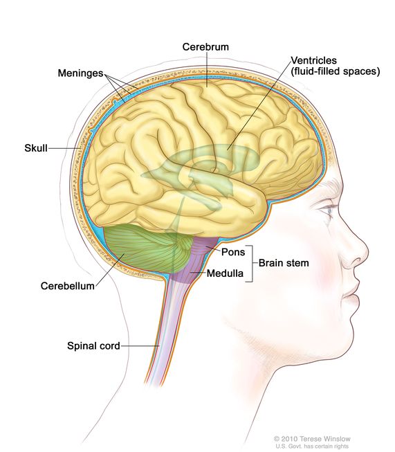
The brainstem is located above the spinal cord and beneath the thalamus and consists of the medulla oblongata, the pons, and the midbrain. Disorders affecting the basal ganglia are still sometimes referred to as extrapyramidal disorders. The basal ganglia are sometimes referred to as the “extrapyramidal system” to differentiate them from the “pyramidal system” (more accurately referred to as the corticospinal tract). The basal ganglia also work in conjunction with the substantia nigra as part of the dopamine circuit, which is damaged in Parkinson’s disease. It scales movement and determines how large, small, fast, or slow a movement needs to be for optimum performance. This part of the brain helps the cerebral cortex execute subconscious, learned movements. A motor command initiated by the cortex is modified and processed within the basal ganglia. The basal ganglia, together with the cerebellum and the motor cortex, are involved with motor control. The cerebral hemisphere (in pink) surrounds the caudate nucleus thalamus (in the center). This image illustrates the left lateral view of the brain and spinal cord, as well as the caudate nucleus and basal ganglia deep in the brain, and a contour of the rest of the thalamus. The names of these three structures are combined in various ways: the caudate nucleus and the putamen together are referred to as the striatum and the globus pallidus and the putamen together are known as the lentiform nucleus. The three masses that compose the basal ganglia are called the caudate nucleus, the putamen, and the globus pallidus. The subcortical structures-the basal ganglia, also known as the extrapyramidal system-are three large masses of cells (ganglia) that lie at the base of the cerebral cortex and surround the thalamus. The thalamus accepts and sifts sensory information and is the part of the brain where sensation is first consciously experienced or felt.

The thalamus is closely integrated with the cerebral cortex and is responsible for the initial processing of all sensory information (except olfaction). Sensation travels to the thalamus from peripheral sensory neurons. The thalamus or “inner chamber” is a small ovoid mass about 3 cm long located at the base of the cerebral hemispheres. The thalamus is the destination of spinothalamic tract-the sensory pathway responsible for processing pain, temperature, and crude touch. The cerebral cortex-especially the frontal areas-is the area of the brain most commonly damaged by stroke. Sensory information flows via bundles of afferent nerves residing in the cortex from the peripheral nervous system for processing. Motor commands flow from efferent nerve fibers originating in the cortex out to the muscles. The cortex originates thoughts and commands and receives information from the periphery and other parts of the brain for processing and interpretation. The cerebral cortex is the thinking and processing part of the brain. New imaging techniques show that the cortex is more extensively interconnected than previously thought. These descriptions derive from early brain research and are no longer considered to be accurate except as a broad overview. The cerebral cortex has historically been divided by function and structure areas into the somatosensory, somatomotor, primary motor, visual, and auditory areas.

The interconnectedness of the nerve cells creates a flexible system, with redundancy that allows recovery of function following injury to the brain.

The cell bodies of the cortex have a high metabolic requirement, using six times more blood than other parts of the brain. The cerebral cortex is highly convoluted and folded, which increases its surface-a phenomenon unique to humans. Axons arising from the estimated 100 billion cell bodies of the cortex run both horizontally and vertically, and each connects with thousands of other neurons, creating a highly complex network. It is the part of the brain most often affected by stroke. The cerebral cortex is a thin layer of nerve cell bodies covering the surface of each hemisphere. The human cortex showing a highly interconnected network of nerve cells.


 0 kommentar(er)
